|
REGULAR ECHINOID TERMINOLOGY |
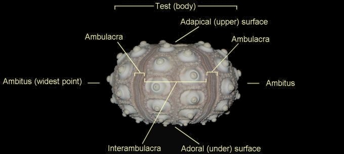
The anatomy and morphology of regular echinoids is strongly influenced by their mode of life (BMNH guide). Their diet is based around material scavenged / predated off the sediment surface, which calls for powerful jaws and a life exposed on the sea-floor. Defence is therefore a priority, and large and elaborate spines are a characteristic feature of the regular echinoids. Mode of life requires that they can roam in any direction, so the body plan is one of radial symmetry (hence 'regular'; i.e. 'circular'); the mouth is located at the centre of the under-surface, and the anus at the centre of the upper-surface.
1). Lateral (side) view of a complete recent cidaroid test (body), lacking its original compliment of spines (x3). The test is divided into five interambulacral areas by five vertical ambulacral columns (BMNH guide).
 A A |
 B
B |
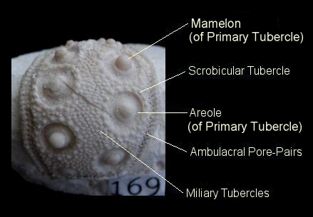
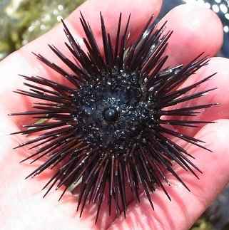
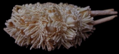
2). (A) Lateral view of an isolated interambulacral segment of Tylocidaris clavigera (x4, adoral end up); cidaroid tests typically disarticulate along the centre of the ambulacral column to leave such segments. The interambulacra are composed of two columns of interlocking pentagonal plates. A primary tubercle is situated at the centre of each interambulacral plate, enclosed within a ring of scrobicular tubercles and an expanse of miliary tubercles. Primary tubercles are comprised of a raised, ball-like mamelon surrounded by a crater-like areole. Different forms of spine are mounted on the various tubecles. The ambulacral pore-pairs act as openings for the tissues of the tube feet; tentacle like projections with a suction-tip, used for clinging and locomotion. (B) Adoral view of a live regular echinoid with it's full compliment of primary spines; from the pages of Discovering Fossils.
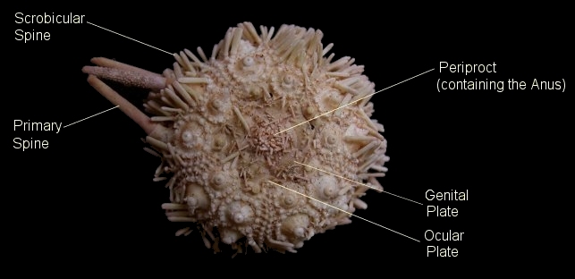
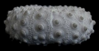
3). Adapical view of a partially-decayed recent cidaroid (x5). The primary spines are mounted on the primary tubercles, and the scrobicular spines are mounted on the scrobicular tubercles. The primary spines form an effective defense against larger predators, with their attachment to the test protected by the scrobicular spines. They also aid in locomotion. Microscopic mounted jaws (pedicellariae) defend the surface of the test against smaller invaders and clear away debris.
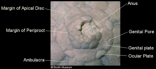
4). Part of a well preserved individual of Phalacrocidaris merceyi retaining the apical disc (x3). Each ambulacral column terminates in an ocular plate, whilst the interambulacral areas terminate in a genital plate. Together, the ocular and genital plates form the oculogenital ring which encloses the periproct; a flexible plated surface which contains the anus (BMNH guide). The oculogenital ring and periproct combined form the apical disc. Reproductive fluids are released through the genital pores. A specialised genital plate (the madreporite) is sieve-like, and is involved in the supply of water to the hydrostatically-operated tube-feet.
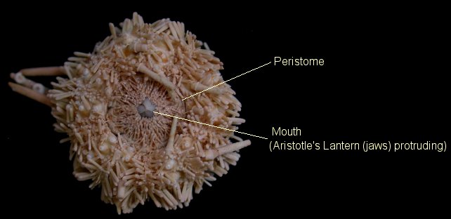
5). Adoral view of a partially decayed recent cidaroid (x5). The peristome is a flexible plated surface which contains the mouth-opening. The complex jaw apparatus (the 'Aristotle's Lantern') (BMNH guide) protrudes through the mouth opening, allowing the cidaroid to feed on material which it passes over. Many different elements make up the lantern, the most recognisable being the ten beak-like hemi-pyramids.
|
|
 B B |
|
|
|
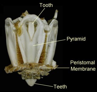
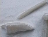
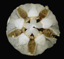
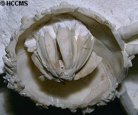
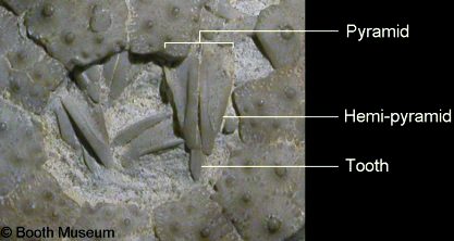
6). The Aristotle's Lantern. Each pyramid is formed of two hemi-pyramids which support an elongate tooth. A lantern is comprised of five pyramids, five teeth, and a number of smaller structures. (A) Recent example of an Aristotle's Lantern (jaw apparatus), x2.5. The membrane of the peristome through which the teeth projected is still attached. (B) View of the beak-like tip of another recent lantern, x2.5. (C) Two fossil hemi-pyramids from the lantern of Tylocidaris clavigera (x2.5), and (D) a hemi-pyramid and a tooth from Hirudocidaris hirudo (x2.5).
 A
A |
 B B |
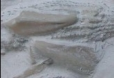
7). Fossilised examples of the Aristotle's Lantern; (A) Test of Hirudocidaris hirudo broken open to expose a complete and articulated lantern (x2.5); (B) Partially articulated fossil lantern of Phymosoma koenigii (x3). (BMNH guide).
 A A |
 B B |
8). Lateral views; (A) Partially decayed recent cidaroid (x5), the area of attachment for the primary spines is clearly well protected by the scrobicular spines; (B) naked fossil test of Phymosoma koenigii (x2).