|
ELASMOBRANCH TERMINOLOGY |
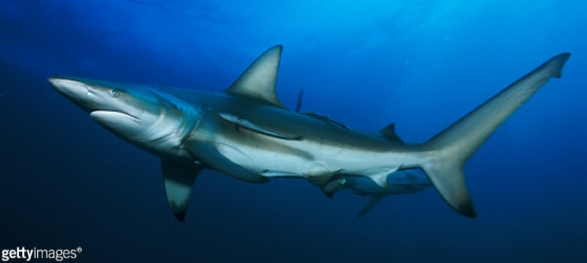
1). A modern Requiem Shark, around 1.5 m long - a cacharhinid with relatives amongst the shark fauna of Late Cretaceous. This individual is sporting a remora/sharksucker fish (echeneid). Image sourced from Getty Images.
The skeletal anatomy of sharks is characterised by a skeleton built entirely of calcified cartilage and a head with amphistylic / hyostylic jaws (upper jaws not fused to the braincase or the hyoid elements which support them). The only materials freely available in the fossil record are the mineralised skeletal elements - teeth (enamel, dentin), placoid scales (dentin) and bony fin spines. The vast majority of fossil specimens are isolated teeth, and classification schemes are necessarily based around tooth morphology. However, tooth form is highly variable both within and between individuals, and also with ontogeny (life stage), often causing great problems for identification of fossils and recognition of taxonomic relationships.
Shark cartilage, except for the vertebral centra, has a distinctive mineralised outer crust of 'tesserae', resembling a mosaic of tessellated tiles. Vertebral centra have a unique form of 'areolar' cartilage with a web-like mineralisation. Both forms of shark cartilage are encountered on occasion in the fossil record, and the distinctive tesserae make this easily recognisable.
 |
 |
 |
| A - LABIAL VIEW | B - SIDE VIEW | C - LINGUAL VIEW |
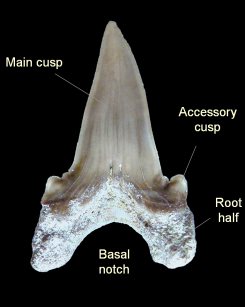
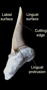
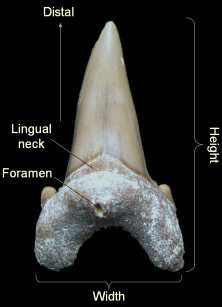
2). Shark tooth terminology - (A) - Labial (outer) surface, faces out from the mouth; (B) Side view; (C) Lingual (inner) surface, faces into the mouth. [Cardabiodon ricki, x3.0, Grey Chalk, Bonchurch, Isle of Wight, Pete King Collection. Images © 2009 Pete King, by kind permission].
Shark taxa are typically defined on a combination of root and crown characters. For example, Cardabiodon, figured here, can be characterised by (amongst other things) generally being very large, with well developed accessory cusps, well separated root halves, a broad, rounded basal notch, a smooth cutting edge, a lingual neck which narrows towards the cutting edges, a flattened labial surface and a convex lingual surface with a clear lingual protrusion.
|
|
 B B |
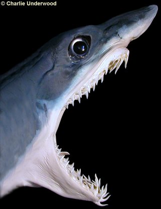
3). Skeletal elements of the shark's head - (A) Right-side view of a stuffed head; (B) Overlay illustrating cartilaginous skeletal elements. [Shortfin Mako (Isurus oxyrinchus), a modern lamniform (lamnid), x0.7, Charlie Underwood, image used by kind permission].
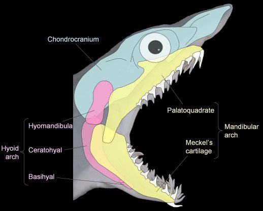
The jaw elements of elasmobranchs (sharks and rays) have amphistylic and hyostylic arrangements. The upper jaw (palatoquadrate) is not fused to the braincase (chondrocranium), the jaws being supported instead by the hyoid elements, which also form the floor of the mouth. Whilst biting, the hyomandibula braces the jaws against the braincase, whilst the palatoquadrate is able to articulate against the braincase. The arrangement allows great flexibility and manoeuvrability of the jaws - the palatoquadrate is often free to move considerably relative to the braincase, often sliding out well away from the ventral surface of the chondrocranium until reaching the end of the "leash" formed by the ligaments that tether it.
The upper jaws (palatoquadrate) and lower jaws (Meckel's cartilage) comprise the mandibular arch. The hyomandibula, ceratohyal and basihyal ('tongue bone') comprise the hyoid arch. The 'arch' terminology reflects the evolutionary origins of the mandibular and hyoid elements as former 1st and 2nd branchial (visceral/gill?) arches respectively.

4). Upper jaws (palatoquadrate) of the Grey Nurse Shark (Carcharias taurus) with the different groupings of teeth and regions of the jaw relating to tooth classification highlighted (x0.6) - terminology after Siverson (1999). The main groupings are the anterior and the latero-posterior teeth, which develop within narrow invaginations (infolds) of the palatoquadrate known as the the anterior and latero-posterior hollows (respectively). Teeth are generated within the hollows and brought forward to join the active set in a conveyor-belt fashion, replacing old teeth which are shed. A shark may generate and shed thousands of teeth in a lifetime.
The anterior and lateroposterior hollows are separated by a small area known as the intermediate bar, whilst the area of junction between the right and left jaw parts is referred to as the symphysial area, also forming an effective bar between the right and left anterior hollows. In some taxa, small teeth develop over the intermediate bar and the in the symphysial area - these are characterised by their irregular form and flattened base to the root.
Individual rows of teeth are referred to as files. The arrangement and relative size of files within the jaws is known as the dental formula, and can be important for classification. The number of files is given with reference to just one jaw set (either the left- or right-hand jaw set). Such species concepts can be very difficult to apply to fossil material.
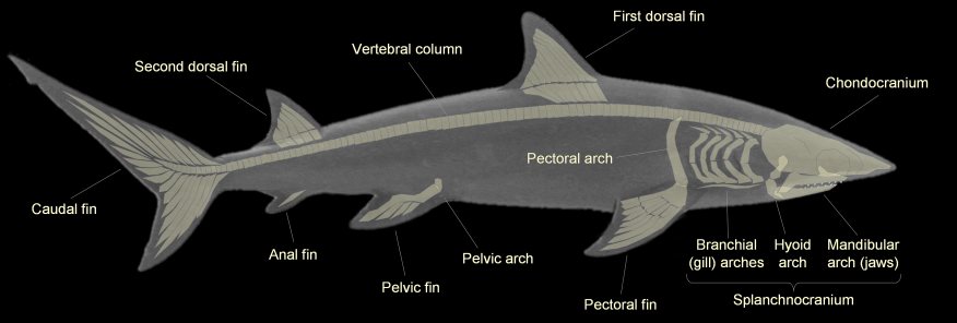
5). Right-side images of a modern shark with the cartilaginous skeletal elements highlighted. The pectoral and pelvic fins are supported by the pectoral and pelvic arches respectively, whilst five pairs of branchial arches support the gills. [Grey Nurse Shark (Carcharias taurus), a modern lamniform (odontaspid) with relatives amongst the shark fauna of the Late Cretaceous, approximately 3 m long].
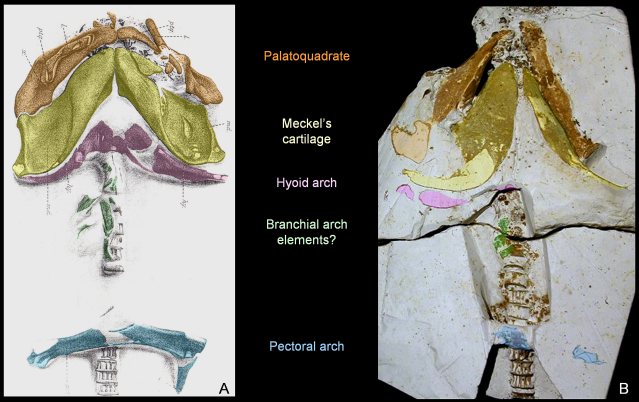
6). Ventral (underside) views of the anterior (front) third of two fossil sharks form the British Chalk, with colour overlays indicating the different skeletal elements. The cartilaginous nature of the skeleton causes such specimens to be extremely scarce in the fossil record, but a small number have been recorded from the British Chalk. [(A) Cantioscyllium decipiens, an orectolobiform (Carpert Shark), figure from Woodward (1902-1912, Plate XXLII, figure 7), x0.7, Grey Chalk, Blue Bell Hill, Burham, Kent, BMNH (British Museum (Natural History) London) P.5890; (B) Heterodontus canaliculatus, a heterodontid (Bullhead Shark), x0.8, White Chalk, Turonian, Malling, near Lewes, East Sussex, Willett Collection, Booth Museum, BMB 007329, by kind permission of John Cooper]
|
|
 B B |
7). Dorsal fin spines - Certain elasmobranch taxa possess bony fin spines supporting their dorsal fins, a relatively 'primitive' elasmobranch character. These are relatively robust elements with a good fossil record. [Modern and a fossil heterodontid (Bullhead Shark) fin spines: (A) Heterodontus francisci (Horn Shark), dorsal fin with distal portion of fin spine protruding vertically; (B) Heterodontus canaliculatus, portion of vertebral column with fin spines (x1.9, Chalk, Booth Museum, BMB 008555), by kind permission of John Cooper)]. Image (A) sourced from Getty Images.
 A
A |
 B
B |
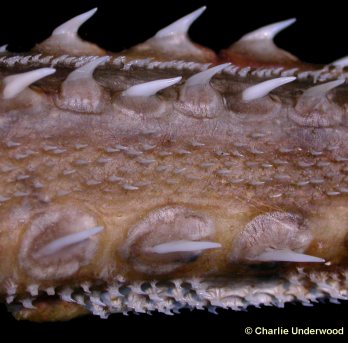
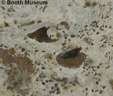
8). Dermal denticles - Elasmobranchs possess tooth-like dermal denticles known as placoid scales. These are made of dentine, and it is likely that sharks teeth originated by modification of denticles around the oral margin. Placoid scales are variably developed amongst shark taxa, sometimes forming a coarse, dense, thorny cover. They have good preservation potential, but are typically small to microscopic and easily overlooked. [Modern and fossil examples of thorny placoid scales - (A) Tail of a modern rajid (skate) x4. Image courtesy of Charlie Underwood; (B) Squatina cranei, a squantinid (Angel Shark), x6.5, Grey Chalk, Clayton, near Brighton, Sussex, Willett Collection, Booth Museum, BMB 007330, by kind permission of John Cooper)].
 A
A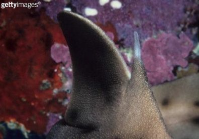 A
A