| FOSSILS | VERTEBRATES | CHONDRICHTHYES | TERMINOLOGY | REFERENCES | LINKS | |
| CHIMAERA (RATFISH) TERMINOLOGY | ||||||
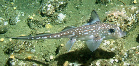
1). Modern Chimaera, the Spotted Ratfish (Hydrolagus colliei). Image sourced from Wikipedia.
Recent Ratfish are largely deep water dwellers and are rather unfamiliar creatures. Having a largely un- or poorly-mineralised cartilaginous skeleton, their fossil record largely comprises isolated elements of their unusual crushing dentition, or dorsal fin spines (venomous in life).
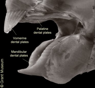
The dentition comprises six dental plates: a pair of mandibular dental plates in the lower jaw; a pair of vomerine dental plates at the front of the upper jaw; and a pair of palatine dental plates at the back of the upper jaw. The vomerine plates couple with the beak at the front of the mandibular plates to form a gripping / biting beak at the front of the jaws. The palatine plates and the rear portion of the mandibular plates form a crushing dentition at the back of the jaws. The inner (gripping / biting / crushing) surfaces are essentially smooth, but with hypermineralised pads or ridges termed tritors.
The form of the dental plates and arrangement of the tritors, particularly for the mandibulars, is commonly used to classify fossil remains. Dental plate form is highly variable within species, and classification is based on general characters.
Chimaera skeletal elements lack the distinctive outer mineralised crust of mosaic-like 'tesserae' seen in sharks and rays.|
|
 B B |
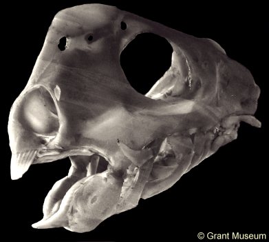
2). Cartilaginous skull of a recent Chimaera: (A) Overview of left side of skull (x1.5); (B) Detail of mouth indicating the arrangement of dental plates which comprise the beak-like jaws. The lower jaw contains a single pair of mandibular dental plates. The upper jaws have a pair of vomerine dental plates at the front, and a pair of palatine dental plates behind (x4). From the collections of the Grant Museum, images used by kind permission.
|
|
 B B |
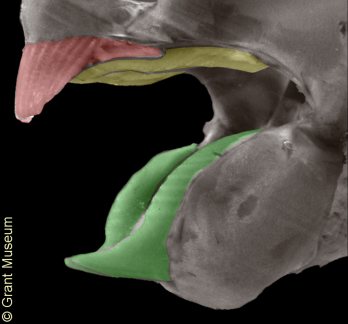
3). A complete set of Chimaera demtal plates from the Chalk (A) and a the jaws of a recent Chimaera (B), with dental plates coloured to demonstrate how the fossil remains fit into the anatomy of the jaws (green - mandibular dental plates; red - vomerine dental plates; yellow - palatine dental plates). (A) Edaphodon sedgwicki x0.55, Lewes, East Sussex, Booth Museum, BMB 009007, by kind permission of John Cooper. (B) Recent Chimaera, x4, from the collections of the Grant Museum, image used by kind permission.
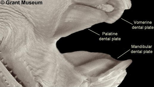
4). Internal view of the left hand side of the mouth of a recent Chimaera (front of head to the right), exposing the biting surfaces of the dental plates - note tritors (see below). From the collections of the Grant Museum, image used by kind permission.
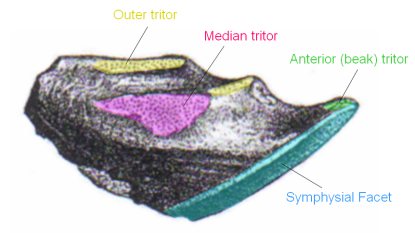
5). Biting surface of a left mandibular dental plate with 'tritors' highlighted. The arrangement of the tritors on the dental plate, the development of the 'beak' at the front of the mandibular plate, the extent of the symphysial facet on the mandibular plate, and overall size are some of the key characters used for classification of fossil Chimaeras. (Ischyodus thurmanni, x1.5, Cambridge Greensand (West Melbury Marly Chalk equivalent), Cambridgesgire?, modified from text figure 56A of Woodward, 1902-1912).
|
|
 B B |
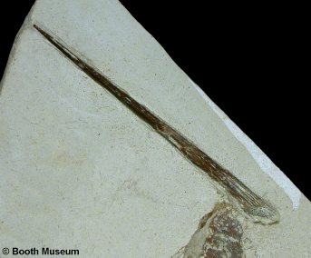
6). Dorsal fin spines: (A) Recent Chimaera displaying prominent fin spine associated with the dorsal fin and (B) fossil dorsal fin spine of Elasmodectes sp. from the Chalk. (A) Modified from image posted by Washington University News.; (B) x2.0, Grey Chalk, West Melbury Marly Chalk, Clayton, near Brighton, West Sussex, Willett Collection, Booth Museum, BMB 007269, by kind permission of John Cooper.
 A
A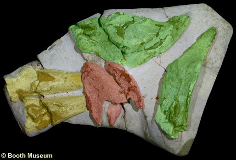 A
A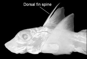 A
A